🌲 Gymnosperm Monograph: Pinaceae Family
A definitive treatise exploring the evolutionary divergence, morphological identification keys, needle fascicle structures, cone architectures, and extreme cold-hardiness adaptations of Pines (Pinus), Spruces (Picea), and Firs (Abies) across boreal and montane biomes.
The conifer division (Pinophyta), specifically within the flagship family Pinaceae, comprises some of the most enduring, structurally massive, and ecologically dominant woody plants on Earth. Dominating the vast circumboreal taiga forest and ascending to the highest timberlines of alpine mountain ranges, conifers exhibit physiological specializations that allow them to endure prolonged sub-zero temperatures, intense desiccation, catastrophic snow-loading, and impoverished, acidic soils. At the forefront of this gymnosperm lineage stand three premier genera: Pinus (Pines), Picea (Spruces), and Abies (True Firs). While untrained observers frequently conflate these evergreen conifers under the generic label of ‘pine trees,’ each genus represents a distinct evolutionary branch defined by rigorous diagnostic characters.
Understanding the anatomical separations among Pinus, Picea, and Abies requires examining leaf attachment biology, vascular needle histology, seed cone orientation and abscission patterns, bark resin duct development, and crown architectural growth forms. Furthermore, these genera exhibit specialized biochemical mechanisms—such as tracheid cell wall lignification, supercooling, and extracellular freezing tolerance—that represent the pinnacle of evolutionary engineering for cold-climate survival. This monograph provides an exhaustive comparative guide to the taxonomy, morphology, and silvicultural ecology of these three iconic gymnosperm genera.
1. Diagnostic Separation: Pinus vs. Picea vs. Abies
Field identification of evergreen conifers relies upon a classic botanical diagnostic triad based on needle arrangement and twig attachment. By memorizing these fundamental morphological distinctions, arborists and naturalists can achieve immediate generic identification in the field within seconds:
1. Genus Pinus (Pines): Needles in Bundles (Fascicles). The defining foliar characteristic of true pines is the arrangement of their long, slender needles into discrete clusters known as fascicles. Each fascicle represents a dwarf shoot (brachyblast) enclosed at its base by a persistent or deciduous sheath of membranous bud scales. The number of needles per fascicle is a primary taxonomic character, typically ranging from two to five needles (rarely one, as in the Single-leaf Pinyon Pinus monophylla, or up to eight). Furthermore, pine cones possess thick, woody scales bearing an elevated central or terminal swelling called the umbo, often armed with a sharp, rigid prickle.
2. Genus Picea (Spruces): Sharp, Square Needles on Peg-like Sterigmata. Spruce needles are borne singly around the twig in a spiraling arrangement. Unlike pines, they are not grouped into fascicles. Each individual spruce needle is attached to the stem atop a distinct, persistent woody peg known as a sterigma (or pulvinus). When a spruce needle desiccates and drops, this woody peg remains permanently affixed to the branchlet, giving dormant spruce twigs a harsh, deeply roughened, rasp-like texture. Furthermore, spruce needles are characteristically four-sided (rhomboid or square in cross-section), allowing them to be effortlessly rolled between one’s thumb and index finger. Spruce seed cones are pendulous (hanging downward from branches) with flexible, papery to leathery scales that drop intact from the canopy upon reaching maturity.
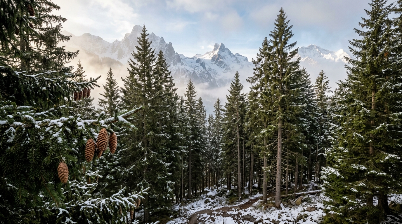
3. Genus Abies (True Firs): Flat, Friendly Needles on Suction-Cup Bases. True fir needles are likewise borne singly, but they are flat, soft to the touch, and frequently display two prominent white, parallel bands of stomatal pores on their abaxial (underside) surfaces (‘flat and friendly firs’). When a fir needle is detached, it leaves behind a smooth, circular, disc-shaped scar resembling a tiny suction-cup footprint, leaving the twig bark entirely smooth and devoid of projecting pegs. Most dramatically, seed cones of Abies are erect (standing strictly upright like upright candles atop upper canopy branches). At maturity, fir cones never fall to the forest floor intact; instead, their cone scales and bracts shatter and disintegrate while still affixed to the crown, leaving a bare, persistent central spike (rachis) standing alone on the branch.
| Diagnostic Feature | Genus Pinus (Pines) | Genus Picea (Spruces) | Genus Abies (True Firs) | Genus Larix (Larches) |
|---|---|---|---|---|
| Needle Arrangement | Bundles (Fascicles of 2-5) | Singly attached, spiraled | Singly attached, 2-ranked/spiral | Clusters of 15-40 on short shoots |
| Twig Attachment Base | Membranous basal sheath | Raised woody peg (Sterigma) | Flat circular disc (Suction cup) | Rosette cushions on dwarf spur |
| Needle Cross-Section | Semicircular to triangular | Four-sided / Rhombic (rolls easily) | Flat / Flattened dorsiventrally | Flat / Triangular, deciduous |
| Cone Orientation | Pendent or horizontal | Pendulous (hanging downward) | Erect (standing upright on upper branches) | Erect, small, persistent on twigs |
| Cone Abscission | Falls intact to forest floor | Falls intact to forest floor | Disintegrates on tree; axis remains | Falls intact or weathers on twig |
| Foliage Phenology | Evergreen (retained 2-10 yrs) | Evergreen (retained 4-10 yrs) | Evergreen (retained 5-12 yrs) | Deciduous (sheds annually in autumn) |
2. Microscopic Needle Anatomy and Cryoprotective Adaptations
The needle-like leaf morphology of conifers represents one of the most sophisticated botanical adaptations to cold, desiccating, and nutrient-poor environments. Unlike the broad laminae of deciduous angiosperms, which present high surface-area-to-volume ratios susceptible to rapid transpiration and frost damage, conifer needles possess a compact cylindrical or flattened cross-sectional geometry that minimizes transpirational water loss.
Examining a pine needle cross-section under high-power optical magnification reveals several specialized histological layers:
- Cuticle and Thick-Walled Epidermis: The outermost layer consists of a dense, heavily lignified epidermis coated with a thick, hydrophobic cuticular wax layer composed of long-chain fatty acids and secondary wax tubules. This cuticular armor reduces non-stomatal cuticle transpiration to negligible levels during winter when the ground is frozen and roots cannot replenish water.
- Sunken Stomatal Crypts: Stomata are arranged in distinct longitudinal lines along the needle surface, recessed deep within sunken pits (crypts). These crypts are frequently filled with porous plugs of epistomatal wax, which create a high-humidity boundary layer above the guard cells, drastically increasing resistance to vapor diffusion while permitting carbon dioxide entry.
- Plicate Mesophyll: The photosynthetic mesophyll tissue beneath the hypodermis is formed of parenchymatous cells with deeply infolded, convoluted cell walls (plicate mesophyll). This morphological folding dramatically amplifies the internal surface area for chloroplast distribution without increasing the physical dimensions of the needle.
- Resin Canals: Lined with secretory epithelial cells, longitudinal resin ducts traverse the mesophyll. These canals synthesize and store oleoresin—a complex mixture of volatile monoterpenes and non-volatile diterpene resin acids that serves as a pressurized chemical defense system against bark beetles and wood-boring larvae.
- Transfusion Tissue and Vascular Bundle: Enclosed within a central endodermis lies transfusion tissue consisting of living parenchyma cells and non-living tracheids surrounding the vascular bundles (one or two bundles, delineating the subgenera Strobus and Pinus).
To survive extreme boreal winters where ambient temperatures plunge below minus 40 degrees Celsius, conifer needles undergo cold acclimation through active biochemical restructuring. They accumulate high concentrations of cryoprotective osmoprotectants—including sucrose, raffinose, stachyose, and proline—alongside specialized cold-regulated (COR) proteins and dehydrins. These molecules depress the cellular freezing point, stabilize cell membranes, and promote controlled extracellular ice nucleation, preventing lethal intracellular ice crystal formation within the living cytoplasm.
3. Cone Biology and Seed Dispersal Strategies
Gymnosperms do not produce true flowers or fruits; their reproductive structures are strobili (cones). In the Pinaceae, plants are monoecious, producing separate pollen-bearing microsporangiate strobili and seed-bearing megasporangiate strobili on the same individual tree. To minimize self-pollination (geitonogamy), pollen strobili are predominantly concentrated in the lower to mid-canopy, whereas seed strobili develop near the highest terminal branch tips, where wind dispersion is maximized.
The morphology of the mature seed cone illustrates profound co-evolutionary relationships with environmental vectors and seed-dispersing wildlife. Conifer seed cones consist of an elongated central axis supporting overlapping spirally arranged scales. Each cone scale is a compound structure comprising an outer sterile bract and an inner fertile seed scale (ovuliferous scale) bearing two winged seeds on its upper (adaxial) surface. In Abies and Pseudotsuga (Douglas-firs), the sterile bracts are often elongated and project prominently beyond the cone scales; in Douglas-fir (Pseudotsuga menziesii), the three-pointed, trident-like bracts projecting past the scales are famously described as the ‘rear legs and tail of a mouse hiding beneath a scale.’
Cone opening mechanisms operate through hygroscopic differential expansion. Conifer cone scales are constructed of two distinct cellular layers: an upper layer of thick-walled, highly lignified sclerenchyma cells that swell minimally with humidity, and a lower layer of cellulose-rich tissue that expands dramatically when wet and contracts when dry. When ambient humidity drops and warm, dry winds blow, the lower tissue contracts while the upper layer remains rigid, forcing the cone scales to flex outward and releasing the winged seeds into atmospheric air currents (anemochory).
Serotiny: Fire Ecology and Pyrogenic Seed Release
In fire-prone forest ecosystems, several pine species have evolved serotinous cones—cones that remain tightly sealed and resin-glued shut on the branches for years or even decades, waiting for heat shock from a wildfire to open. Prominent examples include Lodgepole Pine (Pinus contorta var. latifolia), Jack Pine (Pinus banksiana), and Pitch Pine (Pinus rigida).
The scale tips of serotinous cones are sealed with high-melting-point diterpene resins that require exposure to temperatures between 50 and 65 degrees Celsius to melt. Following the passage of a crown fire that consumes the parent tree canopy, the resin bonds liquefy, allowing the hygroscopic scales to flex open in the dry post-fire convective air. The winged seeds rain down upon the newly cleared, mineral-rich ash bed, which is completely free of competing vegetation, shaded canopy cover, and pathogenic soil fungi. This pyrogenic adaptation ensures immediate, dense post-fire seedling colonization.
4. Cellular Wood Anatomy: Tracheids and Torus-Margo Pits
Conifer timber—commercially classified as ‘softwood’—possesses a cellular histology that is fundamentally simpler and more homogenous than the complex vessel-and-fiber architecture of hardwood angiosperms. Over 90 to 95 percent of the xylem volume in Pinaceae is composed of a single, multifunctional cell type: the tracheid. Tracheids are elongated, narrow, spindle-shaped cells with heavily lignified secondary cell walls that perform both mechanical structural support and water conduction simultaneously.
Because conifers lack the wide, open vessel elements found in oaks and maples, water moves between overlapping tracheids through specialized circular structures known as bordered pits. In conifers, these bordered pits feature an extraordinary microscopic valve called the torus-margo pit membrane. The central portion of the membrane consists of an impermeable, thickened disc called the torus, suspended within the pit chamber by a porous, radiating net of microfibrils known as the margo.
This microscopic valve acts as an autonomous safety mechanism against catastrophic hydraulic failure (cavitation). When freezing temperatures or severe drought stress cause an air bubble (embolism) to form within a tracheid, a severe pressure differential develops across the pit membrane. The high pressure on the embolized side pushes the flexible margo, driving the solid central torus against the circular opening (aperture) of the pit border like a microscopic rubber stopper. This seal aspiration isolates the air bubble, preventing the embolism from spreading into adjacent functioning tracheids and preserving the overall hydraulic integrity of the tree canopy.
6. Conifer Oleoresin Chemistry and Mycorrhizal Symbioses
Beyond structural anatomy, the evolutionary dominance of the Pinaceae across nutrient-starved, acidic podzolic soils is powered by two biochemical alliances: defensive oleoresins and obligate ectomycorrhizal symbioses.
Conifer oleoresin is a complex secondary metabolic solution synthesized within specialized resin canal epithelial cells. It comprises approximately 50 percent volatile monoterpenes and sesquiterpenes (such as alpha-pinene, beta-pinene, and limonene) and 50 percent non-volatile diterpene resin acids (such as abietic and neoabietic acids). Historically harvested as ‘naval stores’ for caulking wooden sailing ships, oleoresin functions in living conifers as a pressurized hydraulic defense system. When a bark beetle (such as the Mountain Pine Beetle, Dendroctonus ponderosae) bores into the phloem, severed resin canals erupt under high hydrostatic turgor pressure, flushing the attacking insect out of the gallery in a sticky mass of resin (‘pitch-out’). Upon contact with atmospheric air, the volatile turpentine fractions evaporate rapidly, leaving the solid rosin acids to oxidize and polymerize into a crystalline, impenetrable seal over the wound, preventing secondary infection by wood-staining fungi (Grosmannia clavigera).
Concurrently, conifers depend upon extensive subterranean partnerships with specialized ectomycorrhizal fungi (EMF), predominantly belonging to the families Boletaceae, Russulaceae, and Suillaceae (such as the slippery jack genus Suillus, which associates almost exclusively with Pinus). The fungal hyphae form a dense mantle around the conifer’s lateral feeder roots and penetrate intercellular spaces in the root cortex to form the Hartig net. This fungal network extends miles into surrounding soil, excreting organic acids and phosphatase enzymes that mobilize insoluble phosphorus and nitrogen from mineral grains, delivering these vital nutrients to the host conifer in direct exchange for photosynthetic sucrose.